Biomedical Image Technologies (BIT)
Dept. Ingeniería Electrónica (Universidad Politécnica de Madrid)

The aim of this activity is to contribute to the design and implementation of high resolution Positron Emission Tomography (PET) scanners. These scanners require to detect and identify pairs of photons coming from the same annihilation positron-electron. The identification of these pairs of almost simultaneous photons is the key to obtain information on the spatial distribution of the radiopharmaceutical emitting positrons. A new research line has been started on Optical Tomography.
The main activities are:
Design of acquisition modules with programmable logicBy continuously sampling the signal provided by the detectors, the acquisition module has to identify the detection of an annihilation photon and to characterize it in terms of energy, spatial position and time of detection. This last feature has to be very accurate as it is the key to identify pairs of detections in coincidence. In this work we have designed an acquisition module based on programmable devices (FPGA). 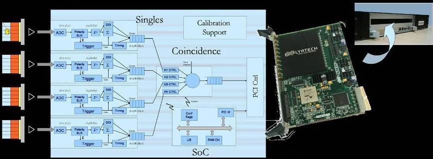
Schematic of PETonCHIP architecture: event and coincidence detection on a single FPGA |
Characterization & simulation of new detectors & architecturesBy means of simulations, the performance of different architectures and detectors has been characterized. Rotating or static architectures have been studied with different number of detectors blocks in order to get the best compromise in terms of cost, spatial resolution and sensitivity. Other detector crystals like CZT are also been simulated to determine the feasibility of their use in PET scanners. This could have a high interest to build combined PET/MRI scanners. A new line has been started on Monte Carlo simulations for turbid media light propagation to assess optical tomography reconstruction. Tomographic reconstructionEfficient 2D & 3D iterative reconstruction algorithms (OSEM) to obtain the best possible images that each scanner can produce have been developed. These algorithms need a system matrix that relates the scanner geometry with the acquired data. We have simulated the system matrix with Monte Carlo techniques. The simulator also allows including in the reconstruction process effects like the probability of detection in the scintillator crystals, probability of Compton scattering and others. |
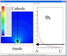
FEM simulation for the computation of weighting potential within a 32 mm thick CZT slab 
18F rat study. FORE + 2D OSEM fast reconstruction |
This material is presented to ensure timely dissemination of scholarly and technical work. Copyright and all rights therein are retained by authors or by other copyright holders. All persons copying this information are expected to adhere to the terms and constraints invoked by each copyright holder. Permission to reprint/republish this material or to reuse any copyrighted component of this work in other works must be obtained from the copyright holders.
![]()

Copyright © 2005-2021 BIT-UPM. Map of web.
Last modified on 17 Jun. 2021 by andres@die.upm.es